
SIGNEL
Case of the Month
History
A 6-year-old girl fell on an outstretched hand and sustained an injury to her right elbow. She presented with gross swelling and deformity of the elbow. On examination, there was no neurovascular deficit. A radiograph revealed Gartland type 3 supracondylar fracture of the right elbow. (Figure 1)
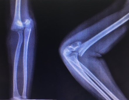
Figure 1
What are your management options at this stage?
She underwent closed reduction and cross Kirschner (K) wires fixation (Figure 2).
Figure 2
Follow-up at three weeks postoperatively in the clinic showed numbness over the little finger and clawing of the little and ring fingers (Figure 3) - (This was not evident in the initial post-operative period).
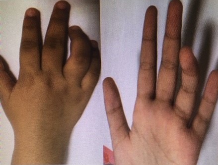
Figure 3
What is the current problem and what initial steps would you take?
Answer: Ulnar nerve neuropathy (delayed)
Initial steps:
Full neurological assessment
Removal of cast/bandages
Removal of wires/metalware
Both wires were removed. However, there was no improvement in the function of the ulnar nerve after over 6 weeks of follow-up.
What would you do at this stage?
Answer: Ulnar nerve exploration and neurolysis
Open exploration and neurolysis of the ulnar nerve was performed 6 weeks after removal of the wires (Figure 4). There was a significant improvement in the function of the nerve noted after two weeks. Full recovery was achieved in 12 weeks (Figure 5).
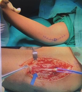
Figure 4
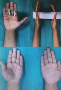
Figure 5
Ulnar Nerve Palsy Following Percutaneous Cross K-wire fixation of Supracondylar Fractures in Children’s Elbows
Introduction
Supracondylar fractures of the humerus are the second most common fractures surpassed only by forearm fractures among upper limb fractures. It is generally agreed that displaced supracondylar fractures (Gartland Type IIb & III) are considered unstable and should be treated operatively by closed reduction and percutaneous K- wire stabilisation as the established gold standard [1]. Biomechanically, the cross Kirschner wire configuration is resilient for any axial rotation, as tested on synthetic bone and human cadaver models [2]. However, medial wire insertion increases the risk of iatrogenic injury to the ulnar nerve due to the anatomical position of the ulnar nerve behind the medial epicondyle.
Discussion
Ulnar nerve injury is a well-known complication of K- wire fixation in the management of supracondylar humeral fractures in children and has been reported up to 20% in the literature [3]. Complete recovery occurs within few days in case of neuropraxia caused by the trauma or during the reduction of the fracture. Lack of full recovery is consistent with significant damage to the ulnar nerve. The K- wire may directly penetrate the wire or the use of power drill can wrap the neural sheath around the wire [4]. In our case, the nerve was found to be trapped by the fibrous tissue behind the medial epicondyle of the humerus. It was apparent that the wire had penetrated the sheath of the nerve causing the formation of adhesions. The release and subsequent transposition of the nerve resulted in full recovery. Numerous variations in technique have been described to minimise the risk of ulnar nerve injury and to achieve sufficient stability [5, 6]. However, it is an important decision when to explore the nerve. We agree with Ikram et al [6], that return of the nerve function is directly proportional to the timing of exploration as happened in our case. Therefore it is advisable to consider early exploration.
- Herring JA. Tachdjian’s Pediatric Orthopaedics: From the Texas Scottish Rite Hospital for Children. Philadelphia: Saunders, Elsevier Inc; 2008:2451–2474
- Woo C, Ho H, Ashik M, Lim K. Paediatric supracondylar humeral fractures: a technique for safe medial pin passage with zero incidence of iatrogenic ulnar nerve injury. Singapore Medical Journal. 2018;59(2):94–97. Doi:10.11622/smedj.2017094
- Huang M, Chehata A, Bonato L. Iatrogenic Ulnar Nerve Palsy Post Supracondylar K-Wire Insertion: Case Report and Review of Literature. J Orthop Rheumatism. 2017;1(2):24-27
- Anuar R, Gooi SG, Zulkiflee O. The Role of Nerve Exploration in Supracondylar Humerus Fracture in Children with Nerve Injury. Malays Orthop J. 2015;9(3):71–74. Doi:10.5704/MOJ.1511.019
- Edmonds EW, Roocroft, JH, Mubarak SJ. Treatment of Displaced Pediatric Supracondylar Humerus Fracture Patterns Requiring Medial Fixation. Journal of Pediatric Orthopaedics. 2012;32(4): 346–351.
- Ikram, MA. Ulnar nerve palsy: a complication following percutaneous fixation of supracondylar fractures of the humerus in children. Injury. 1996;27(5):303–305.


