
Expert Corner
How I do surgery for resistant Congenital Talipes Equinovarus
 Mohammad Arshad Ikram Mohammad Arshad Ikram Seremban, Malaysia Co-author: Gandhi Nathan Solayar
|
Introduction
Idiopathic congenital talipes equinovarus (CTEV), also known as clubfoot, has been a recognised deformity since the time of the Ancient Egyptians and was independently described by both Hippocrates and the Aztecs. Surgical intervention began in the late 1700s with Lorenzís Achilles tenotomy. However, effective soft-tissue releases, osteotomies, and tendon releases were not developed until the late 1800s with the advent of anaesthetic improvements and aseptic techniques. The adoption of the Ponseti technique has resulted in fewer and less-invasive operations for CTEV population, with accompanying improvement in the overall gait pattern and parent satisfaction.
In the setting of resistant CTEV, a variety of further soft-tissue procedures have been described ranging from a limited posterior release to extensive postero-medial surgery. In 1979, Turco described the outcome in feet with resistant CTEV which had failed to respond to non-operative therapy or previous surgical treatment [5]. The aim of soft-tissue release in idiopathic CTEV is to obtain anatomical correction and a mobile well-functioning foot.
Surgery
I prefer to use the classic Turco one stage posteromedial procedure. The child with bilateral resistant CTEV (Figure 1) is placed supine with a surgical pad under the operating foot. A tourniquet is used without exsanguination of the limb which helps in the identification of the neurovascular bundle. An L-shape incision is marked along the medial border of the foot extending from first tarsometatarsal joint up to 6 cm of the distal leg along the medial border of the Achilles tendon.
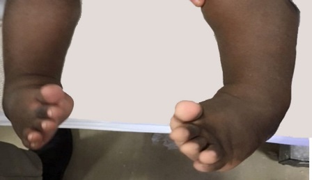
Figure 1: Two-year-old child with bilateral CTEV
An incision is centred below the medial malleolus. After skin incision and subcutaneous tissue dissection, the rest of the exposure is continued bluntly using wet 4x4’s and peanut gauze. The Achilles tendon is dissected off the soft tissue. The paratenon layer is opened and adequate lengthening done using Z-plasty. The neurovascular bundle is identified, dissected free and protected by a vessel rubber loop. Tendons of the flexor hallucis longus and flexor digitorum longus are identified and lengthened using further Z-plasties. The posterior release is completed by incising open the capsule of the ankle and the subtalar joints and releasing the posterior talofibular and the calcaneofibular ligaments. This corrects the equinus deformity and the foot can be placed plantigrade.
Medial release involves exposure and lengthening of the tibialis posterior, release of the knot of Henry, the superficial deltoid ligament, the spring ligament, opening of the capsule of the talonavicular joint, naviculo medial cuneiform joint and the first tarsometatarsal joint. I do release the abductor hallucis muscle if there is persistent forefoot adduction. Full correction is achieved on the operating table (Figure 2).
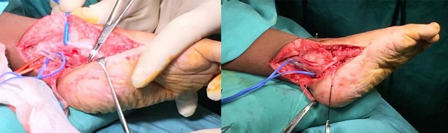
Figure 2: Peri-operative photographs showing soft tissue releases & tendon lengthening with full correction on table
I prefer avoiding the use of K-wire fixation in my practice. The tourniquet is released before closure of the wound. Skin is closed by subcuticular 4/0 absorbable sutures. An above-knee bivalve plaster cast is applied with the foot held in slight under correction. I bring the child back at 2 weeks for suture removal. At that stage, an above-knee plaster cast is applied in full correction with the patient under general anaesthesia for 4 weeks (Figure 3). I advise the parents in the use of a Dennis Brown splint at night for one year (Figure 4).
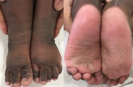
Figure 3: The same patient two months after surgery
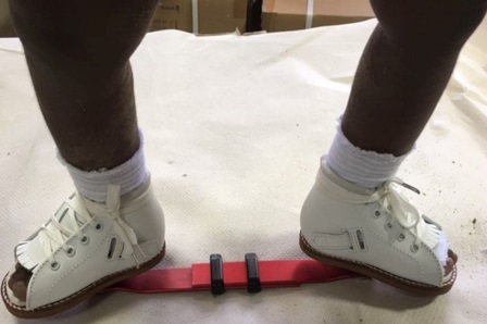
Figure 4: The child in Dennis Brown shoes
- Miedzybrodska Z. Congenital talipes equinovarus (clubfoot): A disorder of the foot but not the hand. Journal of Anatomy.2003; 202 (1): 37-42
- Ponseti.I.V. Observation on pathogenesis and treatment of congenital clubfoot clin orthop.1972; 84: 50-60.
- Morcuende J, Dolan L, Dietz F & Ponseti I. Radical reduction in the rate of extensive corrective surgery for clubfoot using the Ponseti method. Pediatrics.2004; 113: 376-380.
- Duffy, MD,Jose J. Salazar, Lee Humphreys, and Brona C. McDowell. Surgical Versus Ponseti Approach for the Management of CTEV: A Comparative Study Catherine M. J Pediatr Orthop 2013;33:326–332)
- Turco V.J. Resistant congenital clubfoot – One stage posteromedial release with internal fixation. Journal of Bone and Joint Surgery.1979; 61(A) : 805-814.


