
Scientific Debate
Wiberg angle and acetabulum-head index – are these parameters interchangeable in severe cases of Perthes disease?
 A.B. Dzemiantsou A.B. Dzemiantsou Minsk, Belarus |
Summary: We present a correlation analysis to establish the connection between Wiberg angle and acetabulum-head index in severe cases of Perthes disease among the patients treated by triple pelvic osteotomy and conservatively. A direct correlation of Wiberg angle and acetabulum-head index has been revealed before and after treatment. These parameters are interchangeable. This study demonstrated the high efficiency of triple pelvic osteotomy, which resulted in a significant value improvement of Wiberg angle and acetabulum-head index.
Keywords: Perthes disease, triple pelvic osteotomy.
Introduction
Perthes disease cases constitute 25-20% among all hip joint disease cases in children, and in the structure of orthopaedic pathology it varies from 0.2% to 2%. In recent years we have seen an increase in the number of patients followed by an extension of the age spectrum, as well as an increasingly unfavorable progression of the disease. The disease is long-term (up to 3-6 years), and in 26-80% of cases it results in pronounced deformity of the femoral head [2, 3, 4].
Therefore, choosing the correct treatment option to reduce the disabling consequences of the disease and taking into account its various X-ray parameters play an important role [1, 5]. To determine the tactics for the treatment of Perthes disease (conservative or surgical), the degree of the femoral head bone coverage is calculated. Wiberg angle (WA) and acetabulum-head index (AHI) are the parameters that visually reflect the degree of the lateral coverage of the femoral head with the acetabulum. The process of calculating these parameters requires practical experience and is long and laborious.
The study objective was to confirm the presence or absence of correlation between the Wiberg angle and the hollow-head index in severe cases of Perthes disease and, if such correlation existed, to assess its degree.
Materials and methods
In order to restore the anatomy of a hip joint, we performed 53 triple pelvic osteotomies (TPO) in 51 patients (Group 1). We observed 20 children with unfavorable symptoms of the disease and who did not have surgery for various reasons. They were treated conservatively (Group 2). Groups are statistically comparable by the main parameters.
We measured WA and the AHI both before and after (3 years) the treatment in both Groups 1 and 2.
Statistical analysis of the obtained data was carried out using the methods of descriptive statistics. Quantitative indicators are presented as following: average value ± standard deviation. For the purposes of correlation analysis, we used the Spearman rank correlation coefficient. We determined the level of statistical significance of the study at p<0.05.
Results and discussion
The Wiberg angle is the main indicator of the stability of the hip joint in the frontal plane and characterises the degree of the lateral coverage of the femoral head with the acetabulum. According to D. Tonnis [11], in patients aged 9-12 this angle should normally constitute no less than 25°, aged 13-20 - no less than 26-30°.
In early stages of Perthes disease, a decrease of this angle indicates an abnormality in the biomechanics of the joint and an overload of lateral areas of the osteoporotic head, which precedes its deformity.
Evaluating the outcomes of Perthes disease, D. Dickens and M. Menelaus [7] revealed a positive correlation between the degree of WA and the outcome. According to the authors [7], the total degree of WA of less than 20° is characteristic of poor treatment results, 20-25° of satisfactory results, and more than 25° of good results.
The acetabulum-head index is one of the most demonstrative and significant indicators of joint stability, it clearly reflects the inconsistency of the femur head coverage with the acetabulum.
It is calculated from the ratio of the femoral head covered by the acetabular roof to the total width of the head epiphysis multiplied by 100%. According to data from P. Klisic [8], the normal value of AHI is 90%, in case of subluxation it is less than 90%. Moreover, the subluxation is considered to be mild with an index value of 80-89% and severe with a value of 79% or less.
The average WA value before the start of treatment in Group 1 was 5.1 ± 7.05°, and the average AHI value was 66.1 ± 8.12%. During calculations, a pronounced interdependence was identified between these indicators (Spearman coefficient 0.67). A pronounced interdependence (Spearman coefficient 0.67) was also identified in Group 2 before the start of treatment; WA constituted 17.1 ± 7.67° and AHI – 78 ± 10.31%. Three years after TPO, the average values of WA were at 36,6 ± 6,83° and AHI was at 98.9 ± 8.3%, indicating that there is also a pronounced interdependence between these parameters (Spearman coefficient 0.61). Pronounced interdependence (Spearman coefficient 0.67) was also identified in Group 2 three years after the start of treatment; WA was at 13.2 ± 8.5° and AHI was at 73.5 ± 5.7% (Fig. 1, Fig. 2, Table 1).
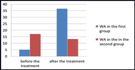
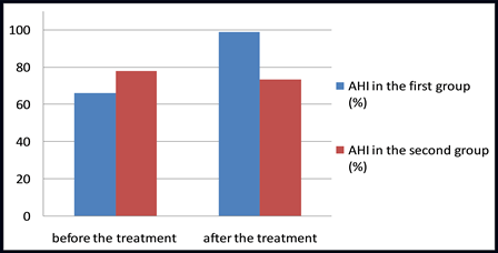
before the treatment | after the treatment | |||
Group of | 5.1±7.05° (WA) | 66.1±8.12% (AHI) | 36.6±6.83° (WA) | 98.9±8.3% (AHI) |
Spearman coefficient 0.67 | Spearman coefficient 0.61 | |||
Control | 17.1±7.67° (WA) | 78±10.31% (AHI) | 13.2±8.5° (WA) | 73.5±5.7% (AHI) |
Spearman coefficient 0.67 | Spearman coefficient 0.67 | |||
The study has shown that in severe cases of Perthes disease, even at the early stages, there is already a progressive deterioration in the parameters characterising the bone coverage of the femoral head with the acetabulum. Conservative treatment does not provide the necessary "coverage" for the femoral head or the stability and does not increase it [6, 10, 11].
We agree with D. Kumar and his co-authors [9] that TPO is the most reasonable and effective surgery to restore "coverage" in severe forms of Perthes disease.
Conclusion
A strong interdependence of WA and AHI before and after the treatment (Spearman coefficient 0,61-0,67) among the patients with severe Perthes disease, treated both surgically with TPO or conservatively is confirmed.
WA and the AHI are interchangeable and interrelated, which will allow orthopedists to use only one of them in order to save time while assessing bone coverage in cases of Perthes disease and while choosing treatment, especially if one uses screening diagnostics or does not have adequate experience in assessing rentgenomentric indicators.
TPO as one of the methods of surgical treatment in severe cases of Perthes disease leads to the improvement of the degree of the femoral head covering, which is reflected in the improvement of WA and AHI.
- D. B. Barsukov. Ortopedo-khirurgicheskoe lechenie detei s bolezn'iu Legga-Kal've-Pertesa: Cand. med. sci. autodiss. 14.00.22. - SPb., 2003. - 28 p.
- M. A. Gerasimenko, A. V. Beletski // Meditsinskiye Novosti Magazine. - 2004. - #3. - P. 40-42.
- A. S. Daurov. Khirurgicheskoe lechenie detei i podrostkov s bolezn'iu Legga-Kal've-Pertesa: Cand. med. sci. autodiss. 14.00.22. – Samara, 1999. – 28 p.
- B. A. Dosanov Usovershenstvovaniye metodov diagnostiki i lecheniya bolezni Pertesa u detei: Cand. med. sci. autodiss. 14.00.22. – Astana, 2009. – 19 p.
- A. I. Korolkov // Ortopedia, travmatologiya i protezirovaniye. - 2008. - #2. - P. 111-120.
- A paired study of Perthes’ disease comparing conservative and surgical treatment / M. Kamegaya [et al.] // J. Bone Joint Surg. [Br]. – 2004. – № 86. – P. 1176–1181.
- Dickens, D. The assessment of the prognosis of Perthes' disease / D. Dickens, M. Menelaus // J. Bone Joint Surg. [Br]. – 1978. – № 60. – Р. 189–194.
- Klisic, P. Treatment of Perthes' disease in older children / P. Klisic // J. Bone Joint Surg. [Br]. – 1983. – № 65. – Р. 419–427.
- Kumar, D. Interlocking triple pelvic osteotomy in severe Legg-Calvé-Perthes disease / D. Kumar, C. Bache, J. O'Hara // J. Pediatr. Orthop. – 2002. – № 22. – P. 464–470.
- Skaggs, D.L. Legg-Calve-Perthes Disease / D.L. Skaggs, V.T. Tolo // Journal of the American Academy of Orthopaedic Surgeons. – 1996. – № 4. – P. 9–16.
- Tonnis, D. Normal Values of the Hip Joint for the Evaluation of X-rays in Children and Adults / D. Tonnis // Clin. Orthop. – 1976. – № 119. – Р. 39–47.


