SICOT e-Newsletter
Issue No. 64 - January 2014
History of Orthopaedics
 Skeletal Injuries and Orthopaedics in Ancient Egypt
Galal Zaki Said
SICOT Distinguished Member â Assiut, Egypt
Egypt had professional doctors as early as the old kingdom, five thousand years ago. There were also specialists in different branches of medicine. Homer c. 800 B.C. remarked in the Odyssey: âIn Egypt, the men are more skilled in medicine than any of human kindâ. Hippocrates, Herophilos, Erasistratus and later Galen studied at the temple of Amenhotep, and acknowledged the contribution of ancient Egyptian medicine to Greek medicine.
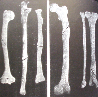
Mummified bodies, skeletal remains and wall paintings have shown us some of the ancient orthopaedic practices. There are many examples of excellently handled fractures of the long bones that had united in perfect alignment. Fractures were treated by splinting with pieces of bark or wood padded with linen. This is particularly impressive in a case of oblique fracture of the femur in an adult, which united without any over-riding (Figure 1).
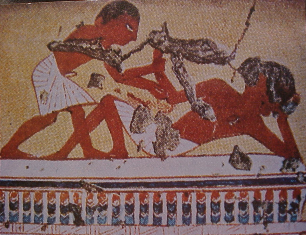
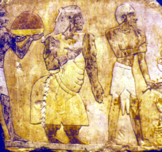
Joint dislocations are depicted in Ipuyâs tomb, Ramses IIâs sculptor, by which a person is setting the shoulder of a prostrate workman, which is similar to the method devised by Kocher for reducing dislocated shoulders, three thousand years later (Figure 2).
Probably the oldest medical document ever written is the Edwin Smith surgical papyrus. The papyrus is a copy of an original which dates to about the 30th century B.C., the time of pyramid building. In this papyrus, forty-eight cases, mostly injuries, were described free from any magic. Descriptions of the patients and their treatment were detailed systematically starting with wounds of the scalp, fracture of the skull exposing the brain, fractures of the neck with paralysis of the arms and legs, fractures of the collarbone and moving down to the extremities. The author, commonly believed to be Imhotep, instructs the treating physician first to listen to the patient's complaint and then to examine him using his eyes and hands. After reaching a diagnosis he makes a declaration: an ailment which I would treat, an ailment which I would contend and an ailment which I would not treat. This formal, structured and logical approach is the basis of our current approach to the patient. Two examples of these cases are cited here:
Infected open fractures with fever were considered grave injuries. The favourite dressing of the wound in the first day was fresh meat (haemostatic). In the following days a dressing of honey (hygroscopic) and oil (to prevent sticking of the dressing) was used unless it was feared it would interfere with drainage. The application of mouldy bread was also practised (in modern days, penicillin was first extracted from moulds).
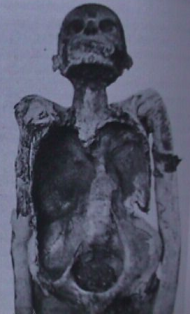
Several cases of tuberculosis of the spine were reported as early as the predynastic time. The most authenticated case is that described in Nesparehan, a priest of Amun. It shows the typical collapse of a dorsal vertebra with angular kyphosis and a big psoas abscess in the right iliac fossa (Figure 3).
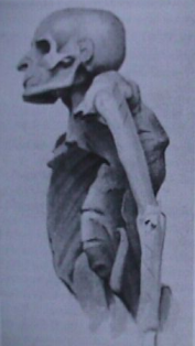
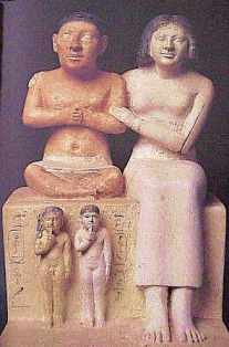
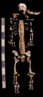
Several examples of congenital anomalies, deformities and hormonal disturbances are present in Ancient Egyptian history. Seneb is a typical example of an achondroplasic dwarf in the 19th dynasty (Figure 4). He held important priesthoods in addition to being overseer of weaving of the palace. Other dwarfs were employed as personal attendants and entertainers. In addition, skeletons of two achondroplasic dwarfs were found in a necropolis of Hierakonpolis in Upper Egypt (Figure 5).
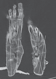
A case of severe bilateral clubfeet is depicted in a Middle Kingdom tomb of Baqt in Beni Hassan. The word djeneb meaning âcrookedâ is written above it. A recent CT scan of the feet of Tutankhamun, the golden Pharaoh of the 18th dynasty, revealed that he suffered from severe varus deformity of his left foot which was also appreciably short (Figure 6). This is probably why the Pharaoh was sometimes depicted using a walking stick.
The Queen of Punt in a bas relief from the temple of Queen Hatshepsut in Dair El Bahari (18th dynasty) raises a diagnostic problem. She is excessively obese, with exaggerated lumbar lordosis, suggesting spondyloptosis or bilateral congenital dislocation of hips (Figure 7).
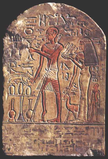
Poliomyelitis was also known at that time as shown in some paintings and sculptures of a doorkeeper, where his leg is wasted, shortened, with an equinous foot and a walking aid is used (Figure 8).
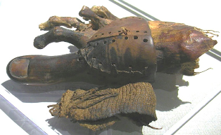
Probably the oldest known prosthesis is that replacing the right big toe in a mummy of a woman found in excavations at the necropolis of Thebes (Figure 9).
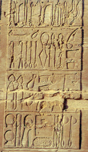
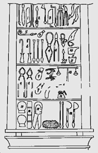
Surgery was evidently advanced in Egypt at the end of the dynastic era as shown by the elaborate surgical instruments engraved in a panel in Kom Ombo Temple (180-47 B.C.) (Figure 10). Two cases of successful amputations, one of arm and the other of leg, were recorded in the literature suggesting the use of the bone saw.
We might never fully grasp the medical and surgical advancement of Ancient Egypt, yet these paintings and sculptures open our imagination to the possible state of Orthopaedics thousands of years ago...
Mummified bodies, skeletal remains and wall paintings have shown us some of the ancient orthopaedic practices. There are many examples of excellently handled fractures of the long bones that had united in perfect alignment. Fractures were treated by splinting with pieces of bark or wood padded with linen. This is particularly impressive in a case of oblique fracture of the femur in an adult, which united without any over-riding (Figure 1).
Figure 1: Fractures of long bones which have united in perfect alignment. Note also the oblique fracture of the femur which has united without any overlap
Joint dislocations are depicted in Ipuyâs tomb, Ramses IIâs sculptor, by which a person is setting the shoulder of a prostrate workman, which is similar to the method devised by Kocher for reducing dislocated shoulders, three thousand years later (Figure 2).
Figure 2: Painting from Ipuyâs tomb in Thebes. A person is setting the shoulder of a prostrate workman
Probably the oldest medical document ever written is the Edwin Smith surgical papyrus. The papyrus is a copy of an original which dates to about the 30th century B.C., the time of pyramid building. In this papyrus, forty-eight cases, mostly injuries, were described free from any magic. Descriptions of the patients and their treatment were detailed systematically starting with wounds of the scalp, fracture of the skull exposing the brain, fractures of the neck with paralysis of the arms and legs, fractures of the collarbone and moving down to the extremities. The author, commonly believed to be Imhotep, instructs the treating physician first to listen to the patient's complaint and then to examine him using his eyes and hands. After reaching a diagnosis he makes a declaration: an ailment which I would treat, an ailment which I would contend and an ailment which I would not treat. This formal, structured and logical approach is the basis of our current approach to the patient. Two examples of these cases are cited here:
- Case 31: Traumatic quadriplegia: âIf you examine a man having dislocation in a vertebra of his neck, should you find him unconscious of his two arms and his two legs, while his phallus is erected⦠and urine drops from his member without his knowing itâ¦â ââ¦it is a dislocation of a vertebra of neck extending to his backboneâ¦, an ailment for which nothing is doneâ.
- Case 35: Fracture of the clavicle: âIf you treat a man for a break in his collarbone and you find his collarbone shortened and out of alignment with respect to its companion, an ailment I will treat. Place him prostrate on his back with something folded between his shoulder blades; you should spread out with his two shoulders to stretch apart his collarbone until break falls in its placeâ.
Infected open fractures with fever were considered grave injuries. The favourite dressing of the wound in the first day was fresh meat (haemostatic). In the following days a dressing of honey (hygroscopic) and oil (to prevent sticking of the dressing) was used unless it was feared it would interfere with drainage. The application of mouldy bread was also practised (in modern days, penicillin was first extracted from moulds).
Several cases of tuberculosis of the spine were reported as early as the predynastic time. The most authenticated case is that described in Nesparehan, a priest of Amun. It shows the typical collapse of a dorsal vertebra with angular kyphosis and a big psoas abscess in the right iliac fossa (Figure 3).
Figure 3: Nesparehan: a priest of Amun, 21st dynasty: Pottâs disease of the spine with typical deformity and a big right psoas abscess
Several examples of congenital anomalies, deformities and hormonal disturbances are present in Ancient Egyptian history. Seneb is a typical example of an achondroplasic dwarf in the 19th dynasty (Figure 4). He held important priesthoods in addition to being overseer of weaving of the palace. Other dwarfs were employed as personal attendants and entertainers. In addition, skeletons of two achondroplasic dwarfs were found in a necropolis of Hierakonpolis in Upper Egypt (Figure 5).
 |
 |  |  |  |
|
Figure 4: Seneb, achondroplasic dwarf, with two children replacing his normal legs
|
 |  |  | Figure 5: Skeleton of achondroplasic dwarf, from Hierakonpolis, Upper Egypt |
A case of severe bilateral clubfeet is depicted in a Middle Kingdom tomb of Baqt in Beni Hassan. The word djeneb meaning âcrookedâ is written above it. A recent CT scan of the feet of Tutankhamun, the golden Pharaoh of the 18th dynasty, revealed that he suffered from severe varus deformity of his left foot which was also appreciably short (Figure 6). This is probably why the Pharaoh was sometimes depicted using a walking stick.
Figure 6: CT scan of Tutankhamunâs feet showing shorter left foot, the seat of varus deformity
The Queen of Punt in a bas relief from the temple of Queen Hatshepsut in Dair El Bahari (18th dynasty) raises a diagnostic problem. She is excessively obese, with exaggerated lumbar lordosis, suggesting spondyloptosis or bilateral congenital dislocation of hips (Figure 7).
Figure 7: Queen of Punt, from Dair El Bahari Temple, 18th dynasty
Poliomyelitis was also known at that time as shown in some paintings and sculptures of a doorkeeper, where his leg is wasted, shortened, with an equinous foot and a walking aid is used (Figure 8).
Figure 8: Ruma, a doorkeeper from the 19th dynasty, is portrayed on his funeral stella with a grossly wasted and shortened leg accompanied by an equinus deformity suggesting poliomyelitis
Probably the oldest known prosthesis is that replacing the right big toe in a mummy of a woman found in excavations at the necropolis of Thebes (Figure 9).
Figure 9: Prosthesis of the right big toe made of wood and leather
Surgery was evidently advanced in Egypt at the end of the dynastic era as shown by the elaborate surgical instruments engraved in a panel in Kom Ombo Temple (180-47 B.C.) (Figure 10). Two cases of successful amputations, one of arm and the other of leg, were recorded in the literature suggesting the use of the bone saw.
Figure 10: Surgical instruments, Kom Ombo Temple: (1) knives; (2) drill; (3) saw; (4) forceps or pincers; (5) censer; (6) hooks, and so on
We might never fully grasp the medical and surgical advancement of Ancient Egypt, yet these paintings and sculptures open our imagination to the possible state of Orthopaedics thousands of years ago...
International Orthopaedics â © Springer-Verlag Berlin Heidelberg 2013 10.1007/s00264-013-2183-z â Published online: 17 November 2013