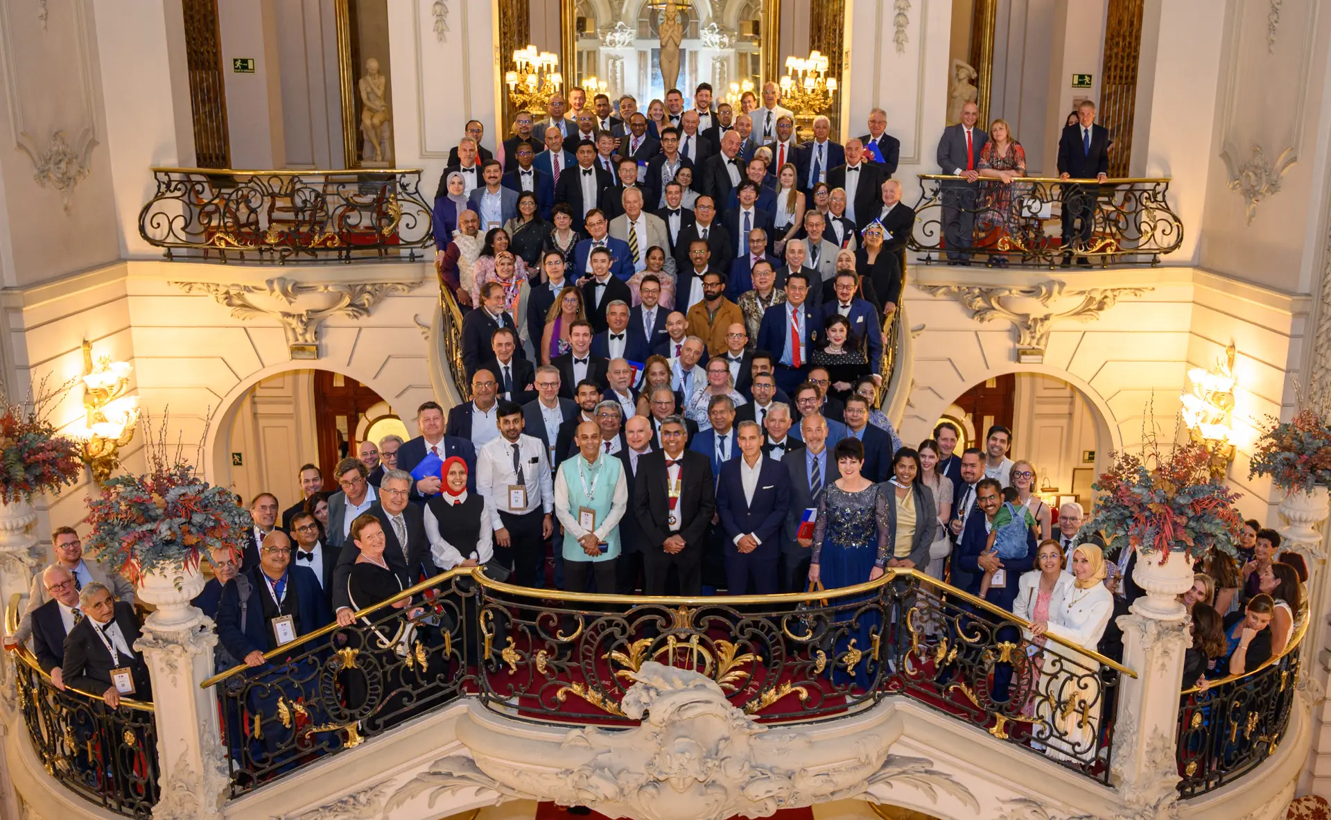
Organisation
Voting Rights
Please find below the list of National Representatives who are eligible to vote in the upcoming elections in Kyoto.
Please find below the list of National Representatives who are eligible to vote in the upcoming elections in Kyoto.

Registration for the prestigious 2026 SICOT Diploma Examination is now open. This internationally recognised qualification benchmarks orthopaedic knowledge and surgical competence, offering candidates a valuable credential that demonstrates excellence within the global orthopaedic community.

The SICOT 2026 World Congress is heading to Kyoto, Japan — and registration is now open. Join orthopaedic and traumatology specialists from around the world for what promises to be an unmissable scientific and professional gathering.
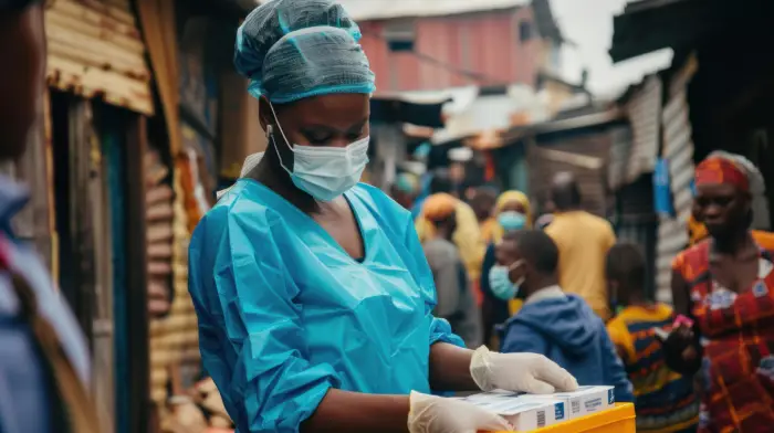
As conflict, displacement and natural disasters continue to affect millions worldwide, humanitarian crises are placing unprecedented pressure on healthcare systems. This article explores the global challenges facing vulnerable populations and highlights the vital role of international cooperation, medical aid and humanitarian action in alleviating suffering.
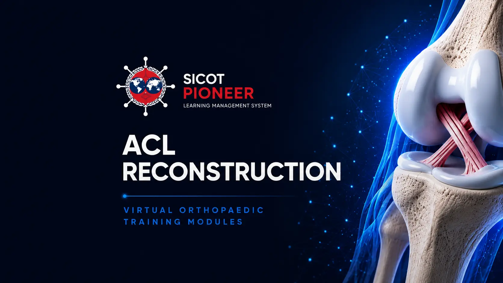
The 2026 PIONEER Virtual Training Course on ACL Reconstruction is open, offering orthopaedic surgeons access to cutting-edge surgical education through SICOT’s online learning platform. An ideal opportunity to sharpen technique and expand clinical knowledge remotely.
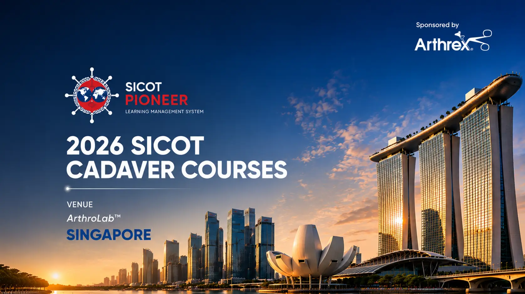
SICOT’s 2026 Cadaver Courses are now available for registration, providing hands-on surgical training in a controlled environment. These intensive practical sessions are designed to enhance surgical proficiency and are open to orthopaedic professionals at all career stages.