SICOT e-Newsletter
Issue No. 69 - August 2014
History of Orthopaedics
History and evolution of intramedullary nailing
Ahmed F. Seifeldin & Ahmed Khedr
SICOT Associate Members - Cairo, Egypt
From the use of intramedullary wooden pegs by Aztecs (1) to the use of ASLS, intramedullary nailing has passed through several stages of development. There have been periods of great enthusiasm and days when the operation was described as a âdaring operationâ according to Time magazine (2).
In this article we will go through the development of intramedullary nails and highlight the important turning points in its history.
In the 16th century, the anthropologist Bernardino de Sahagunand and other conquistadors in America reported on the treatment of delayed union of long bone fractures by Aztec healers with the use of intramedullary wooden rods (1).
During the 19th century and early 1900s most of the work was directed towards nonunions and not fresh fractures. Ivory pegs which reabsorb in the human bodies were used (3,4). In the 1890s, Gluck of Germany presented an intramedullary locking ivory device (5). A similar principle was reported, nearly at the same time, by the Norwegian Nicolaysen for proximal femoral fractures (6). In 1912, Ernest Hey Groves of the United Kingdom was the first to use metallic rods as intramedullary fixation devices. Following the manipulation of open femoral fractures during World War I, he described intramedullary nailing as an easy technique to allow for fixation through very small incisions without additional damage of the periosteum (7). However, due to high infection rates, Groves was nicknamed âseptic Ernieâ and his method didnât propagate. Later in 1917, Hoglundof in the United States reported on the use of autologous cortical bone graft for intramedullary splinting (8). In the 1920s, Smith-Petersen introduced a nail to fix subcapital femoral fractures (9). Later, the Rush brothers from Rochester, Minnesota, described the use of metallic pins placed in the medullary canal to treat fractures of the proximal ulna and proximal femur (10). Meanwhile, Gerhard Küntscher in Hamburg developed intramedullary nailing with the use of a metallic nail, starting the âKüntscher eraâ.
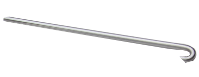
Fig. 1 - Diagram showing a rush nail
Born in Germany in 1900, Küntscher's earliest interest in intramedullary fixation was influenced by his work with the Smith-Petersen. He conducted several cadaveric and animal studies during development of his âmarrow nailâ where he designed the V-shaped stainless steel nail that was inserted with an antegrade method. By 1947, he had performed 105 cases using the V-shaped nail with the help of Finnish surgeons as he was sent to the northern Finnish front because his work was not well appreciated in Germany at that time. By the late 1940s, Küntscher modified the design of the nail to become cloverleaf (11,12).
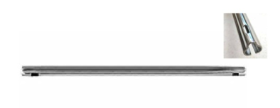
Fig. 2 - Diagram showing Küntscher femoral nail with a cloverleaf cross-section
The use of the Küntscher nail was first described in the United States in a 1945 Time magazine article entitled âAmazing Thighboneâ. It described the treatment of an American prisoner of war by German doctors. The American doctors called the rod technique âa daring operationâ and wondered how their German colleagues inserted it without dangerously cutting down blood supply and without introducing infection (2,12). In the 1950s, Fischer in Germany started to use intramedullary reamers to increase the contact area between the nail and host bone, to improve stability of the fracture (13). However, it took another decade with Küntscherâs introduction of flexible reamers for the concept to take hold. Later on the concept of using interlocking screws to increase stability emerged in 1953 by Modny and Bambara. They presented the multi-hole locking intramedullary nail at a 90-degree angle (14). Also in 1953, Herzog modified the straight Küntscher nail by adding a proximal angle to allow the insertion of the nail above the tibial tubercle to avoid injury to the articular surface (15).
Although intramedullary nailing âslowed downâ in the 1960s due to increased enthusiasm for compression plating, this period witnessed the birth of cephalomedullary nails (16). In 1967, Zickel used a proximally locked nail for the fixation of subtrochanteric femoral fractures.
The Zickel nail had a separate hole for a separate nail that could be placed through the lateral cortex of the proximal femur into the neck and head (17).
In the 1970s, the complications associated with compression plating renewed the interest in intramedullary nailing, especially with the development of image intensification techniques which allowed many surgeons to adopt closed nailing. A wide variety of intramedullary nails were used (ÎÎ and Grosse-Kempf). The concept of using unreamed nails for open fractures was introduced (16).
In the 1990s and early 21st century, the use of intramedullary nails expanded to include proximal and distal fractures and some intraarticular fractures. The gamma nail was designed to fix subtrochanteric fractures together with the retrogade supracondylar nail GSH (Green-Seligson-Henry). New titanium nails were introduced. Slotted cloverleaf designs were being replaced by non-slotted designs to provide greater torsional rigidity. Recon nails, expert tibial nails and ASLS (angular stable locking screws) were developed (16).
Intramedullary nailing has passed through several stages in its development. Although the work of Küntscher was met by scepticism early on, intramedullary nails are by far the standard treatment of diaphyseal fractures and their use in other types of fractures is expanding.
References:
Ahmed F. Seifeldin & Ahmed Khedr
SICOT Associate Members - Cairo, Egypt
From the use of intramedullary wooden pegs by Aztecs (1) to the use of ASLS, intramedullary nailing has passed through several stages of development. There have been periods of great enthusiasm and days when the operation was described as a âdaring operationâ according to Time magazine (2).
In this article we will go through the development of intramedullary nails and highlight the important turning points in its history.
In the 16th century, the anthropologist Bernardino de Sahagunand and other conquistadors in America reported on the treatment of delayed union of long bone fractures by Aztec healers with the use of intramedullary wooden rods (1).
During the 19th century and early 1900s most of the work was directed towards nonunions and not fresh fractures. Ivory pegs which reabsorb in the human bodies were used (3,4). In the 1890s, Gluck of Germany presented an intramedullary locking ivory device (5). A similar principle was reported, nearly at the same time, by the Norwegian Nicolaysen for proximal femoral fractures (6). In 1912, Ernest Hey Groves of the United Kingdom was the first to use metallic rods as intramedullary fixation devices. Following the manipulation of open femoral fractures during World War I, he described intramedullary nailing as an easy technique to allow for fixation through very small incisions without additional damage of the periosteum (7). However, due to high infection rates, Groves was nicknamed âseptic Ernieâ and his method didnât propagate. Later in 1917, Hoglundof in the United States reported on the use of autologous cortical bone graft for intramedullary splinting (8). In the 1920s, Smith-Petersen introduced a nail to fix subcapital femoral fractures (9). Later, the Rush brothers from Rochester, Minnesota, described the use of metallic pins placed in the medullary canal to treat fractures of the proximal ulna and proximal femur (10). Meanwhile, Gerhard Küntscher in Hamburg developed intramedullary nailing with the use of a metallic nail, starting the âKüntscher eraâ.
Fig. 1 - Diagram showing a rush nail
Born in Germany in 1900, Küntscher's earliest interest in intramedullary fixation was influenced by his work with the Smith-Petersen. He conducted several cadaveric and animal studies during development of his âmarrow nailâ where he designed the V-shaped stainless steel nail that was inserted with an antegrade method. By 1947, he had performed 105 cases using the V-shaped nail with the help of Finnish surgeons as he was sent to the northern Finnish front because his work was not well appreciated in Germany at that time. By the late 1940s, Küntscher modified the design of the nail to become cloverleaf (11,12).
Fig. 2 - Diagram showing Küntscher femoral nail with a cloverleaf cross-section
The use of the Küntscher nail was first described in the United States in a 1945 Time magazine article entitled âAmazing Thighboneâ. It described the treatment of an American prisoner of war by German doctors. The American doctors called the rod technique âa daring operationâ and wondered how their German colleagues inserted it without dangerously cutting down blood supply and without introducing infection (2,12). In the 1950s, Fischer in Germany started to use intramedullary reamers to increase the contact area between the nail and host bone, to improve stability of the fracture (13). However, it took another decade with Küntscherâs introduction of flexible reamers for the concept to take hold. Later on the concept of using interlocking screws to increase stability emerged in 1953 by Modny and Bambara. They presented the multi-hole locking intramedullary nail at a 90-degree angle (14). Also in 1953, Herzog modified the straight Küntscher nail by adding a proximal angle to allow the insertion of the nail above the tibial tubercle to avoid injury to the articular surface (15).
Although intramedullary nailing âslowed downâ in the 1960s due to increased enthusiasm for compression plating, this period witnessed the birth of cephalomedullary nails (16). In 1967, Zickel used a proximally locked nail for the fixation of subtrochanteric femoral fractures.
The Zickel nail had a separate hole for a separate nail that could be placed through the lateral cortex of the proximal femur into the neck and head (17).
In the 1970s, the complications associated with compression plating renewed the interest in intramedullary nailing, especially with the development of image intensification techniques which allowed many surgeons to adopt closed nailing. A wide variety of intramedullary nails were used (ÎÎ and Grosse-Kempf). The concept of using unreamed nails for open fractures was introduced (16).
In the 1990s and early 21st century, the use of intramedullary nails expanded to include proximal and distal fractures and some intraarticular fractures. The gamma nail was designed to fix subtrochanteric fractures together with the retrogade supracondylar nail GSH (Green-Seligson-Henry). New titanium nails were introduced. Slotted cloverleaf designs were being replaced by non-slotted designs to provide greater torsional rigidity. Recon nails, expert tibial nails and ASLS (angular stable locking screws) were developed (16).
Intramedullary nailing has passed through several stages in its development. Although the work of Küntscher was met by scepticism early on, intramedullary nails are by far the standard treatment of diaphyseal fractures and their use in other types of fractures is expanding.
References:
- J., Farill. Orthopedics in Mexico. 1952, Vol. 24, pp. 506-512.
- Medicine: Amazing thighbone. Time. March 12, 1945, Vol. XLV., 11.
- F., konig. Uber die Implantation von Elfenbein zum Ersatz von Knochen und Gelenken. Nach experimentellen und klinischen Beobachtungen. Beitr Klin Chir. 85, 1913, Vol. 85, pp. 91-114.
- H., Bircher. Eine neue Methode unmittelbarer Retention bei. Arch Klin Chir. 1866, Vol. 34, pp. 410-422.
- T., Gluck. Autoplastic transplantation. Implantation von Fremdkörpern. Berl Klin Wochenschr. 1890, Vol. 19.
- J., Nicolaysen. Lidt on Diagnosen og Behandlungen av. Fr.colli femoris. Nord Med Ark. 1897, Vol. 8, p. 1.
- EW, Hey Groves. On the application of the principle of extension to comminuted fractures of the long bone, with special reference to gunshot injuries. Br J Surg. 1914, Vol. 2, 7, pp. 429-443.
- EJ., Hoglund. New method of applying autogenous intramedullary bone transplants and of making autogenous bone-screws. Surg Gynecol Obstet. 1917, Vol. 23, pp. 715-759.
- Smith-Petersen MN, Cave EF, Van Gorder GW. Intracapsular fractures of the neck of the femur. Treatment of internal fixation. Arch Surg. 23, 1931, pp. 715-759.
- Rush LV, Rush HL. A technique for longitudinal pin fixation of certain fractures of the ulna and of the femur. J Bone Joint. 1939, Vol. 21, pp. 619-626.
- G., Küntscher. Die Marknalung von Knochenbruchen. Langenbecks. Arch Klin Chir. 1940, Vol. 200, pp. 443-455.
- Vecsei V, Hajdu S, Negrin LL. Intramedullary nailing in fracture treatment: history, science and Küntscher's revolutionary influence in Vienna, Austria. Injury. 2011, Vols. 42Suppl 4:S1-5.
- Fischer AW, Maatz R. Weitere Erfahrungen mit der Marknagelung. Arch Klin Chir. 1942, Vol. 203, p. 531.
- Modny MT, Bambara J. The perforated cruciate intramedullary nail: Preliminary report of its use in geriatric patients. J Am Geriatr Soc. 1953, Vol. 1, pp. 579-588.
- K., Herzog. Nagelung der tibiaschaftbrüche mit einem starren nagel. Arch für Clin Chir. 1953, Vol. 276, pp. 227-229.
- Bong MR, Koval KJ, Egol KA. The history of intramedullary nailing. 2006, Vol. 64, 3 & 4.
- RE., Zickel. A new fixation device for subtrochanteric fractures. Clin Orthop Relat Res. 1967, Vol. 54, pp. 115-123.
  Â